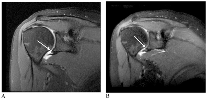
Figure 7.

Humeral Avulsion of the Anterioinferior Glenohumeral Ligament. A) Oblique coronal proton density 2D-FSE image with fat saturation. B) Oblique coronal 3D-FSE-Cube image. The shoulder pathology can be seen in any oblique orientation using the isotropic 3D-FSE-Cube acquisition sequence (arrows, A, B).
