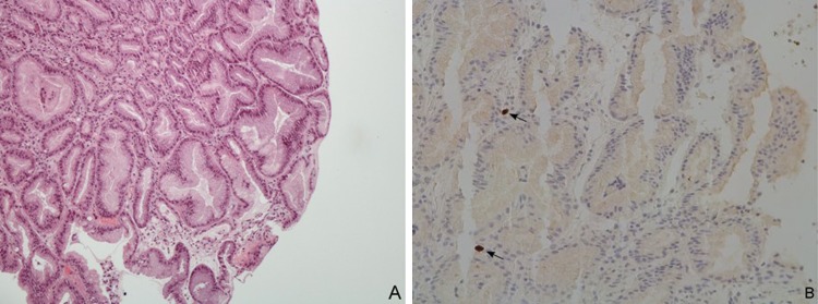
Figure 3.

Light microscopy of gastric biopsy specimens collected from fundus and body. (A) Hyperplasia and cystic dilatation of gastric pits (H&E x200). (B) Cytomegalovirus intranuclear inclusions (arrows) in epithelial cells from gastric body, detected by immunohistochemistry (x200).
