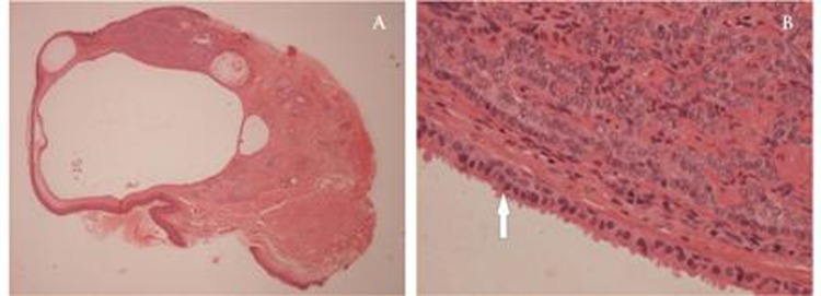
Figure 2.

Low power light microscopy reveals an intradermal loculated cyst with an adjacent solid tumour towards the top of the section, extending to the excision margin (H&E x25). The cyst lining consists of a double layer of cells with ‘decapitation’ of the cytoplasm on the luminal border (arrow), characteristic of an apocrine hidrocystoma on high power. Adjacent cords of basaloid cells with peripheral pallisading, characteristic of a basal cell carcinoma, are seen in the upper part of the section (H&E x400).
