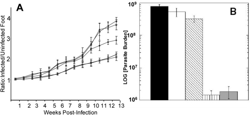
Figure 4. NKT Cells are Required for Increased Protection Induced by αGalCer.

To verify the role of NKT cells in the αGalCer enhancement of protection induced during DNAp36 priming, groups of NKT deficient BALB/c mice were vaccinated and challenged with Leishmania major: -■-, PBS-NK-T (control) -●- pCINeoDNA and WR-Luc (vector control); -▲-, DNAp36 + VVp36; -∗-, p36DNA+αGalCer + VVp36; also shown is the course of infection for control -◆-, PBS WT-BALB/c mice; (DNAp36 versus DNAp36+ αGalCer p=0.45).
B) Evaluation of parasite burdens at 13 weeks post-infection.
 , BALB/c (WT) PBS (control);
 , NK-T PBS (Control);
 , NK-T pCINeoDNA and WR-Luc (vector control);
 , NK-T DNAp36 + VVp36;
 , NK-T DNAp36+αGalCer + VVp36. (DNAp36 versus DNAp36+ αGalCer P=0.65) These results are representative of 2 independent experiments. It should be noted that this experiment was performed together with the experiment shown in Figure 5. Hence an enhanced protection was observed for BALB/c mice vaccinated with p36DNA+αGalCer.
