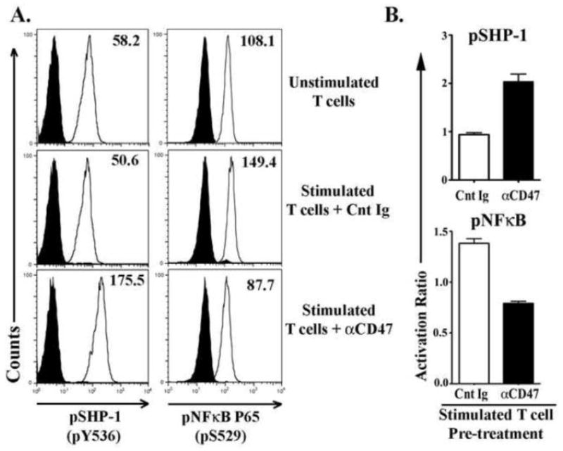
Figure 4. CD47 engagement in control T-cells inhibits TCR induced NFκB activation.

Freshly isolated T-cells from healthy control donors were pre-incubated with αCD47 antibody or an isotype matched nonspecific control immunoglobulin (Cnt Ig) for 30 minutes, then stimulated with plate-bound αCD3 Ab for 3 hours. Unstimulated T-cells served as controls for background phosphorylation levels. Cells were harvested and stained for intracellular pSHP-1 (pY536) and pNFκB p65 (pS529) and assessed by flowcytometry. (A) Histograms showing SHP-1 and NFκB phosphorylation levels in T-cells. (B) Graphs showing the activation ratio of SHP-1 and NFκB in αCD47 or Cnt Ig treated TCR-stimulated T-cells. Activation ratio has been calculated as- [Mean fluorescence intensity (MFI) in treated T-cells/MFI in unstimulated T-cells]. Data represented as Mean ± SEM, n=3.
