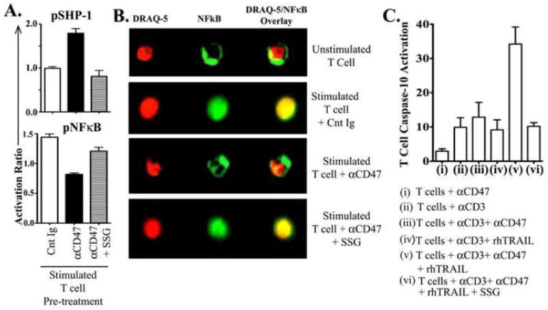
Figure 5. Inhibition of SHP-1 by the pharmacological inhibitor, Sodium Stibogluconate (SSG), prevents CD47-induced NFκB deactivation and rescues T-cells from TRAIL-induced apoptosis.

Freshly isolated T-cells from healthy control donors were pre-incubated either with a control immunoglobulin (Cnt Ig), or αCD47 antibody (Ab) or αCD47 AB + 15 μg/ml SSG for 30 minutes, then stimulated with plate-bound αCD3 Ab for 3 hours. (A) Activation ratio of intracellular pSHP-1 and pNFκB p65 were assessed as described above. Activation ratio has been calculated as [Mean fluorescence intensity (MFI) in treated T-cells/MFI in unstimulated T-cells]. Data represented as Mean±SEM, n=3. (B) Cells were stained for intracellular NFκB p65 (total protein) and nuclear dye DRAQ-5 to assess nuclear/cytoplasmic localization of NFκB by Amnis Image-cytometry. Data are representative of three experiments with similar results. (C) Freshly isolated T-cells were cultured with (i) 10μg/ml αCD47 Ab only, or (ii) immobilized αCD3 antibody alone, or (iii) αCD3 + αCD47 Ab, or (iv) αCD3 + rhTRAIL [1 μg/ml], or (v) αCD3 + rhTRAIL + αCD47 Ab, or (vi) αCD3 + rhTRAIL + αCD47 Ab + 15 μg/ml SSG for five days. Active Caspase-10 levels were determined as previously described. Data shown as Mean±SEM, n=3.
