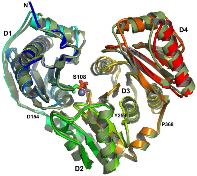
Figure 4.
NMR chemical shift-refined homology model of PMM/PGM superimposed upon the crystal structure. The solution NMR-enhanced model is colored the spectrum of the rainbow from blue at the N-terminus to red at the C-terminus. The crystal structure of the free state (PDB accession code 1K35)16 is green. The side chain of phosphoSer108 is drawn at the base of the catalytic cleft where its Oγ is one ligand of the metal ion16 (gray and presumed to be zinc). Loop junctions between domains are marked with residue numbers.

