
Figure 4.
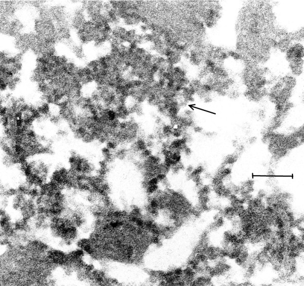
(Mag. 150K). EL case #19940. An image of VLP in the cytoplasm of a neuron. An image taken at low magnification (not shown) revealed large numbers of lipofuscin granules in the cytoplasm of a neuron, displacing the VLP to the periphery of the cell. Figure 4 is a high magnification of an area in the cytoplasm of the same cell, in which VLP are embedded in putative virus factory (arrow). Bar = 150 nm.