Figure.

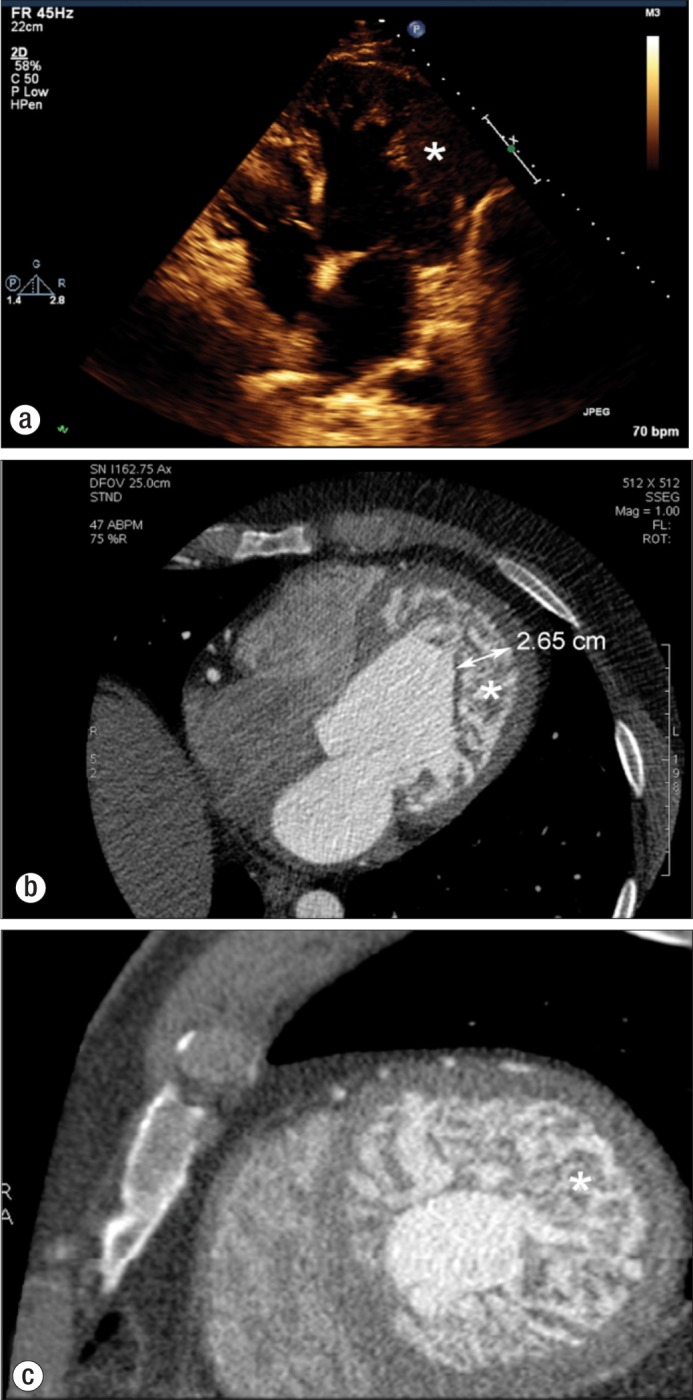
(a) Echocardiographic four-chamber view demonstrating thickened endocardium with numerous sinusoids (asterisk). Computed tomographic images in (b) an axial plane and (c) a short-axis plane clearly demonstrate a noncompacted endocardium (asterisk) that is at least twice the thickness of the compacted myocardium.
