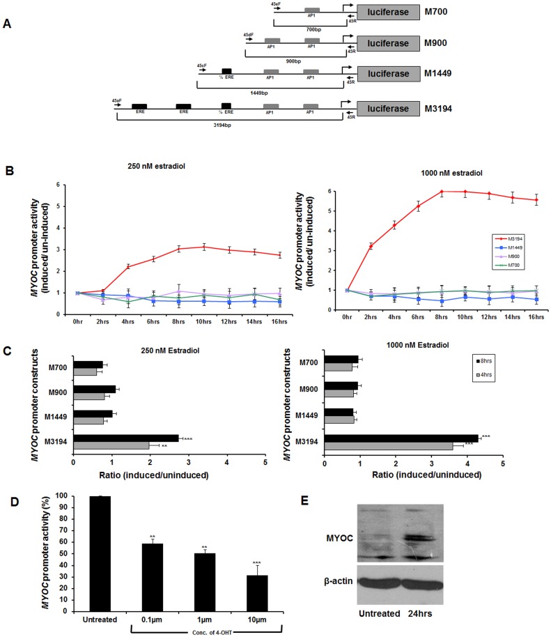
Figure 3. Functional evaluation of putative EREs in MYOC promoter.
A: Serial constructs of MYOC-promoter region containing ERE and AP1 sites cloned in promoter less PGL3 basic vector. Black solid arrows indicate the forward and reverse primers used to amplify the inserts for subcloning. Also, the alphanumeric nomenclature of the constructs corresponds to the first initial of myocilin (M) followed by the size of the insert in base pairs. B : Luciferase activity in extracts from RPE cells transfected with the clones containing MYOC constructs and treated with 17β estradiol (250 nM or 1000 nM). C : Ratio of luciferase activity in cell extracts between induced and uninduced RPE cells for all 4 serial constructs upon dose (250 nM and 1000 nM) and time (4 hrs & 8 hrs) dependent treatment of 17β estradiol. The time points were taken based on the previous experiment in Panel B. D : The M3194 construct was transfected in RPE cells and subjected to increasing amount of 4-hydroxy tamoxifen (4-OHT; 17β estradiol competitor) treatment followed by luciferase assay. A gradual decrease in MYOC promoter activity was observed with increasing amount of 4-OHT. E : Significant upregulation of endogenous myocilin with 17β estradiol treatment in HTM cell. (**p-value<0.001, ***p-value<0.0001). Three independent replicates were performed for all the experiments described here.

