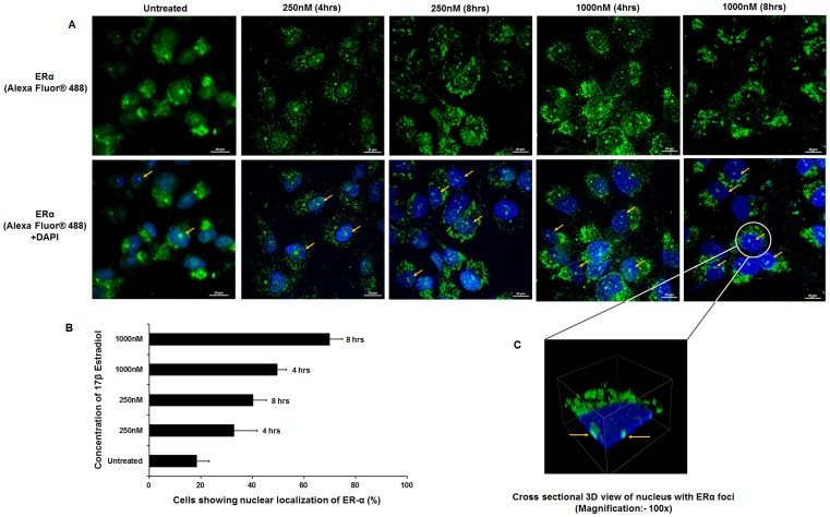
Figure 4. Nuclear localization of ERα on 17β estradiol treatment in HTM cells.
A: Confocal images of HTM cells upon dose (250 mM & 1000 mM) and time (4 hr and 8 hr) dependent treatment with 17β estradiol. Cells were stained with human specific ERα-antibody followed by Alexa Fluor® 488 labeled anti-rabbit secondary antibody (Upper panel). For all conditions, corresponding superimposed image with DAPI are given (Lower panel). Arrows point to the cells where nuclear localization of ERα was observed. B : Histogram showing the percentage of HTM cells with ERα localized in the nucleus upon treatment with 17β estradiol in a dose and time dependent manner. Each experiment was done in triplicate. C : Cross sectional 3D view of nuclear localization of ERα in HTM cell is shown [Scale bar: 20 µm].

