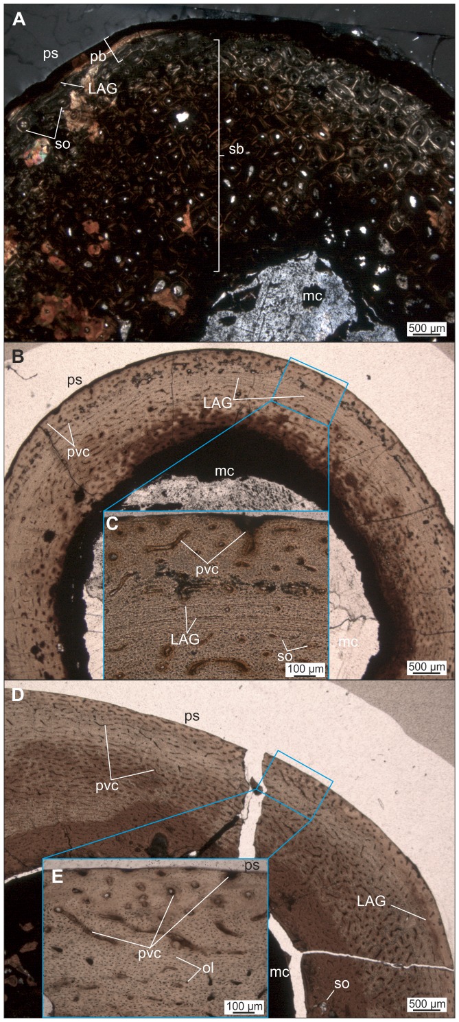
Figure 11. Thin sections of different long bones of Mochlodon suessi.
Histological features show that both, adult (A) and juvenile (B–E) ontogenetic stages are represented in the sample. A. Diaphyseal cross section of tibia PIUW 2349/35. B–C. Diaphyseal cross section of radius PIUW 3517 with close up (C) of the outermost cortical microsturcture. D–E. Diaphyseal cross section of femur PIUW 2349/III with close up (E) of the outermost cortical microsturcture. Abbreviation: pb, primary bone. For further abbreviations see Figure 9.

