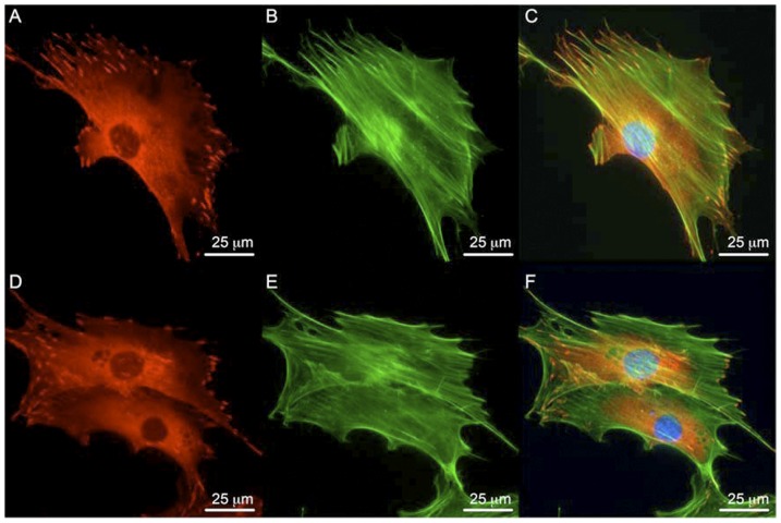
Figure 1. Focal adhesion formation and actin cytoskeleton in wild-type and FAK−/− osteoblasts.
(A–C) FAK+/+ osteoblasts form focal adhesions (red) as shown by vinculin staining and display prominent actin fiber formation (green) as shown by phalloidin staining. (D–F) FAK−/− osteoblasts also exhibit actin fiber and focal adhesion formation. Panels C and F show merged images. DAPI nuclei stain in blue. Magnification = 60×. Scale bar represents 25 µm.

