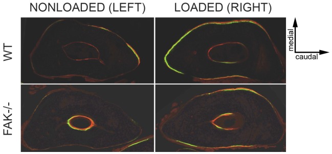
Figure 7. Transverse cross-sections at ulnar midshaft in nonloaded (left) and loaded (right) forearms in WT (top) and FAK−/− (bottom) mice given calcein (green) and alizarin (red) fluorochrome bone labels at 4 and 11 days, respectively, after the first day of loading.
In response to applied mechanical loading, most new bone is formed on the medial and lateral surfaces where bending strains are highest. Note the appearance of double bone labels (green and red) on the medial and lateral surfaces of the ulna, as well as on the rostral surface, in the loaded ulna (top, right) compared to the nonloaded internal control (top, left) where very little bone formation is observed. The loaded ulna (bottom, right) in FAK−/− mice exhibit bone formation on the medial and lateral ulnar surfaces, but much less new bone formation, in terms of percent mineralizing surface, is observed relative to the nonloaded ulna (bottom, left). Sections are representative of the response observed for WT and FAK−/− mice. Magnification = 10×.

