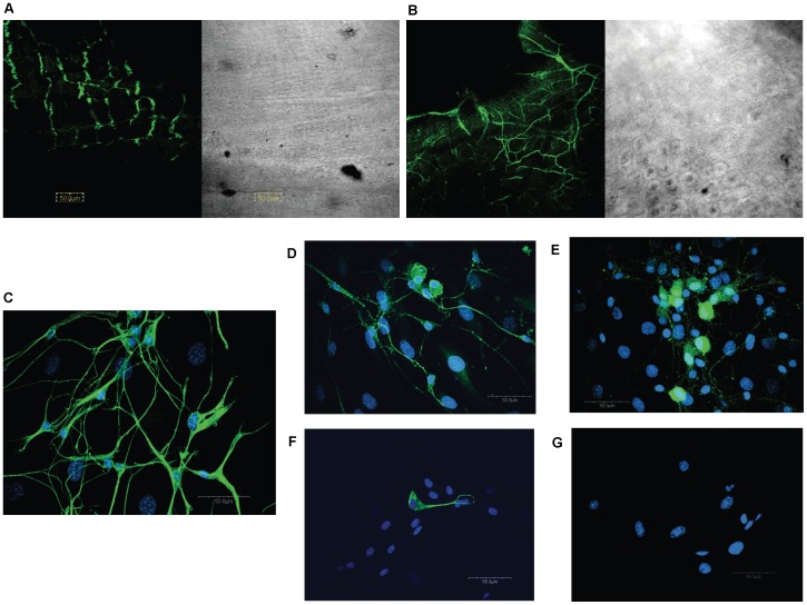
Figure 1. Immunohistochemical characterization of enteric neurons in the mouse.
Confocal microscopy revealed neuronal-specific βIII-tubulin (Abcam, rabbit, 1∶1,000) staining in whole mount ileal longitudinal muscle (A) and circular muscle (B) preparations from the mouse. Cells isolated from longitudinal muscle/myenteric plexus (LMMP) preparations contain neurons (C; βIII-tubulin, Abcam, rabbit, 1∶1,000) that stain postiviely for calbindin (D) (Chemicon, rabbit, 1∶1,000) and calretinin (E) (Swant, rabbit, 1∶2,000). Glia (F) were visualized with the glia-specific marker GFAP (Chemicon, mouse, 1∶500). Antibodies were visualized via appropriate goat secondary antibody Alexa 488 (green, Molecular Probes, 1∶1,000)0. Nuclei were visualized using Hoescht 33342 (blue, C–G, 1 µg/ml). No staining was seen when primary antibody was omitted (G).

