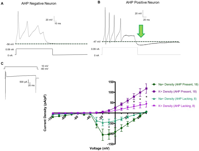
Figure 2. Cultured myenteric neurons are comprised of two electrophysiologically distinct populations.
In current clamp mode (A & B), all neurons displayed action potentials upon current injection of 0.09 nA. Upon cessation of current stimulation, neurons either returned to their original resting membrane potential (A), or dipped below baseline in a slow after-hyperpolarization (AHP, arrow, B). AHPs have an average magnitude of −7.41±0.98 mV, a duration of 212.27±24.98 ms, and a τ = 98.47±13.12 ms. In voltage clamp mode (C), sharp inward spikes of Na+ current followed by a sustained outward K+ current were readily apparent (inset). Current density – voltage relationships of Na+ and K+ currents in both AHP negative and AHP positive neurons showed that AHP positive had significantly greater current densities as determined by two-way ANOVA (* = p<0.05).

