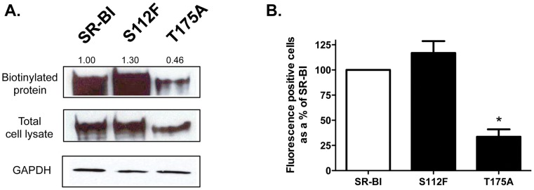
Figure 1. Cell surface expression of wild-type and mutant SR-BI receptors.
(A) COS-7 cells expressing wild-type or mutant SR-BI receptors were assessed for cell surface expression following incubation with NHS-LC-Biotin as described in Materials and Methods. Immunoblot analyses of biotinylated SR-BI at the cell surface (from ∼150 µg of total lysate) (top panel) and in 20 µg of total cell lysate (middle panel) are shown using an antibody directed against the C-terminal cytoplasmic domain of SR-BI. GAPDH was detected as a loading control (bottom panel). The numbers above the top panel represent cell surface receptor expression by densitometry analysis (where SR-BI = 100%). Data are representative of 3 independent experiments. (B) Surface expression of wild-type or mutant SR-BI receptors in COS-7 cells was assessed by flow cytometry using an antibody directed against the extracellular domain of SR-BI. Data are expressed as a % of SR-BI expression following subtraction of empty vector values. Data are the average of 12 independent transfections.

