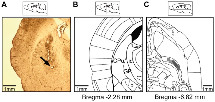
Figure 1. Verification of electrode placement in the Globus Pallidus of rats.
A: a 60 micron slice showing electrode placement in a rat GPe following electrolytic lesion. B: Appropriate coronal section from atlas (Bregma: −2.28 mm; [47]). C: Recording sites marked (grey rectangle) for all animals on a planar rat atlas slice (Bregma: −6.82 mm; [47]).

