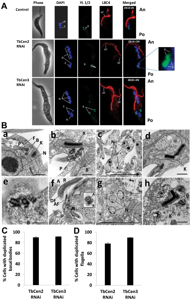
Figure 3. Effect of ablation of centrins on cell shape and organelle number.
A: Effects on the duplication and segregation of basal bodies, kinetoplasts and nuclei in TbCen2 and 3-depleted cells. The cells were stained with DAPI for the nuclei and kinetoplasts, YL1/2 for the basal bodies and L8C4 for the flagella. Note the RNAi induced cells are large and pleomorphic in shape with multiple organelles with more than one flagellum and the new flagella are of detached type (middle and lower panels) compared to the control cell with organelles in single number with one attached flagellum (top panel). Scale bar, 5 µm. B: Electron microscopy of centrin-depleted cells. (a) A typical control cell in this particular section shows single flagellum, pair of basal bodies, kinetoplast and nucleus. (b–d) TbCen2-depleted cells with (b) multi basal bodies and the kinetoplast with enlarged size compared to the control in ‘a’, (c) multi nuclei and (d) abnormal kinetoplast. (e–h) TbCen3-depleted cells with (e) multi basal bodies, (f) multi flagella. Inset is the cross-section image of a detached flagellum displaying the normal axoneme (with 9+2 microtubule structure) and the paraflagellar rod, (g) multi nuclei and (h) an abnormal kinetoplast. Scale bars, 500 nm (a, b, d–f and h) and 2 µm (c and g). C & D: Bar graphs showing the percent of T. brucei procyclic cells with duplicated basal bodies (C) and flagella (D). The cells were analyzed on day 3 after induction for TbCen2 RNAi cells and day 2 for TbCen3 RNAi cells. Data represent the means ± SD of three independent experiments. For each TbCen2 and 3 RNAi studies, over 140 cells were manually counted and analyzed. F, flagellum; B, basal body; K, kinetoplast; N, nucleus; P, flagellar pocket, A, axoneme; R, paraflagellar rod; AF, attached flagellum; DF, detached flagellum; An, anterior; Po, posterior.

