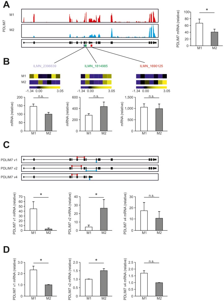
Figure 6. Detection of alternative splicing in human macrophages.
(A) Summarized expression of all PDLIM7 transcripts in human M1- and M2-like macrophages. Left, representative images of sequencing reads across genes expressed in human macrophages as described in Fig. 4. Right, RPKM values for PDLIM7 by RNA-seq in M1- and M2-like macrophages. (B) Expression of PDLIM7 as determined by microarray analysis using 3 different probes recognizing different parts of the PDLIM7 transcripts as depicted in (A). (C) Upper panel: representation of the 3 different mRNA transcripts from Refseq. Lower panel: abundance of the different transcripts as determined using Cuffdiff. (D) qPCR for the 3 different mRNA transcripts from Refseq in human M1- and M2-like macrophages. Splice variant specific primers depicted in red and blue. Data are representative of three experiments (RNA-seq), seven experiments (microarray analysis) or at least ten experiments (qPCR; mean and s.e.m.), each with cells derived from a different donor. *P<0.05 (Student’s t-test).

