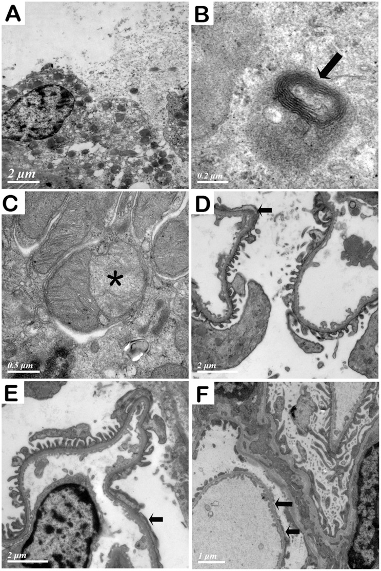
Figure 5. Ultra structural examination of the kidney from the rats with organic solvent treatment.
A. Brush border and cytoplasm of tubular epithelial cell were dropped and epithelial cells were disintegrated. B. An autophagolysosome (arrow) in a tubular epithelial cell. C. Mitochondria in a tubular epithelial cell were degenerated. The inner membrane of a mitochondrion was stripped from outer membrane, forming a space in between (asterisk). D, E, F. Foot process of podocytes after exposure to GDF at 8, 10 and 12 weeks, respectively. Segmental foot process fusion at 8 and 10 weeks was indicated with arrows. Partial foot process was stripped from the glomerular basement membrane at 12 weeks (arrows).

