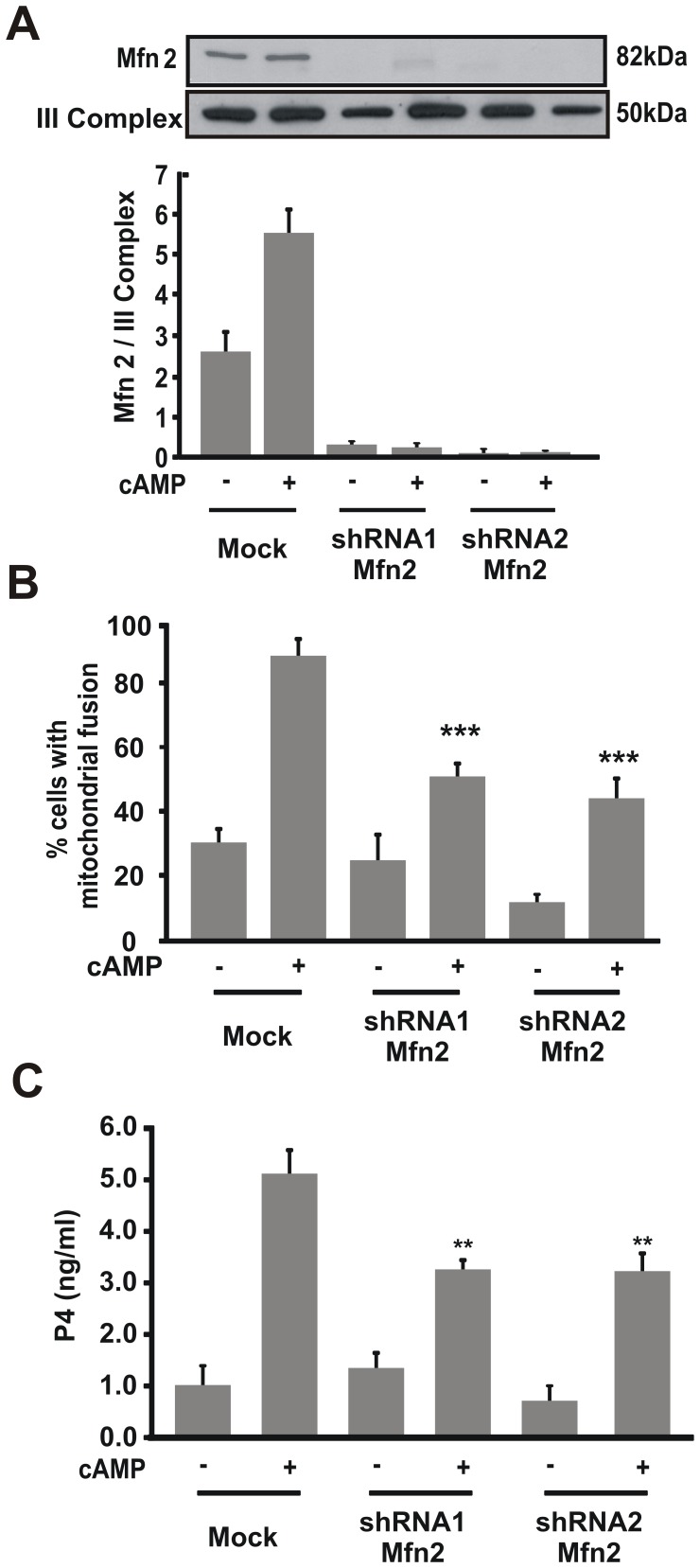
Figure 7. Mfn2 protein is necessary for steroid synthesis.
MA-10 cells were transfected with a plasmid containing different shRNA Mfn2 (shRNA1 or shRNA2). After 48 h, cells were stimulated with 8Br-cAMP (0.5 mM) for 1 h. A. Isolated mitochondrial proteins were obtained and western blotting was performed. Membranes were sequentially blotted with anti-Mfn2 and anti-III Complex antibodies. An image of a representative western blot is shown. For each band, the OD of the expression levels of Mfn2 protein was quantified and normalized to the corresponding III Complex protein. The relative levels of Mfn2 protein are shown. B. Cells were fixed and scored as previously described. Quantitative analysis of mitochondrial fusion is shown. The results are expressed as the means ± SEM of three independent experiments. **P<0.01 vs. cAMP mock. C. P4 levels were determined by RIA in the incubation media. Data represent the means ± SEM of three independent experiments and expressed as ng/ml. **P<0.01 vs. 8Br-cAMP mock.

