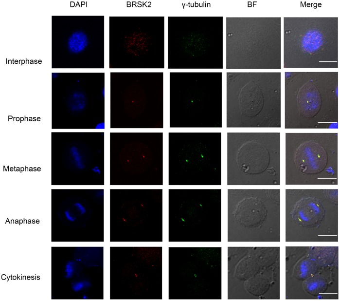
Figure 1. BRSK2 co-localizes with the centrosomes during mitosis.
Synchronously growing HeLa cells were fixed and stained as described in “Materials and Methods”. This figure shows representative images of dividing cells captured in different channels for BRSK2 (red), γ-tubulin (green), and DAPI (blue). All images were captured with an Olympus FluoView FV1000 confocal fluorescence microscope and a 60x oil immersion objective lens. BF: bright field. Scale bar, 10 µm.

