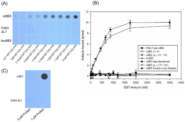
Figure 2.
Analysis of the binding of the D3D4 domain of ankyrin to cdb3. His-tagged cdB3 was incubated for 2h with increasing concentrations of a GST fusion construct of the D3D4 domain of ankyrin and then captured onto Co2+-NTA agarose beads. After washing 3x with binding buffer, bound protein was eluted and quantitated (A) by immunoblot analysis using an antibody to D3D4 ankyrin, or (B) by measuring GST activity in the eluent (see Methods). (C) Alternatively, GST- D3D4 was incubated for 2 hrs with either wild type or a mutant cdB3 lacking loop 2, and the complexes were captured on GST-Sepharose beads, washed 3x with binding buffer and evaluated for cdb3 content by dot blot analysis using an anti-cdB3 antibody. kdcB3 - kidney cdB3; cdB3 ΔL1 – cdB3 lacking loop 1; cdB3 Δ1–50 – cdB3 lacking loop residues 1–50. Error bars correspond to S.D., where n=2.

