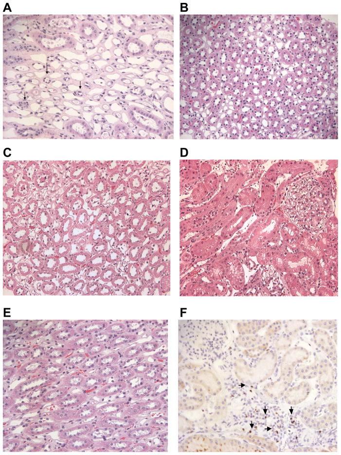
Figure 4. Biopsies of kidney allografts.
Biopsy of G774 (A) demonstrating significant atrophic tubules (arrows) among normal appearing distal tubules but no evidence of tubulitis, changes likely due to drug toxicity rather than acute rejection. Biopsy of H309 (B) demonstrating normal appearing tubules without evidence of tubulitis. Biopsy of H128 (C and D) demonstrating normal appearing tubules, collecting ducts and glomeruli without evidence of tubulitis. Biopsy of G918 showing normal distal tubules and no tubulitis (E) and stained for FoxP3 showing positively stained cells (arrows) proximal to tubules (F). Photomicrographs were taken at 200x magnification.

