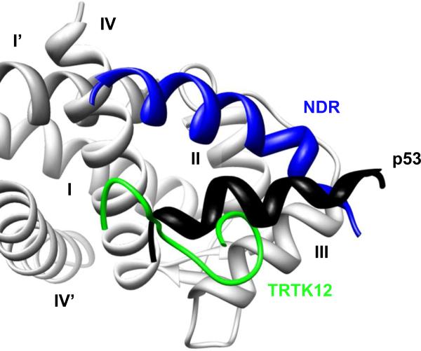
Figure 4.
S100 Proteins Interacts with Peptide Targets through a Promiscuous Binding Site. A ribbon cartoon representing the converged NMR ensemble for the S100B protein interacting with the TRTK12 (green, PDB: 1MQ1), p53 (black, PDB: 1DT7), and NDR (blue, PDB: 1PBS) peptides in the presence of calcium. Only the binding site of one S100B monomer (light gray, Helices I-IV) is shown. Although all three peptides interact with similar amino acid residues in the binding pocket of S100B and with the same stoichiometry, each peptide binds in unique orientation with a distinct structure.

