
Figure 1.
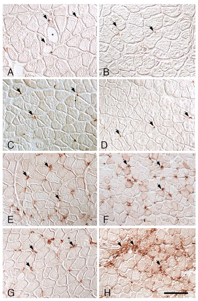
CD68+ macrophage distribution in soleus muscles. A, C, E, G: Wild-type muscles. B, D, F, H: IL-10 null mutant muscles. A, B: Ambulatory control muscles showing CD68+ macrophages (arrows) in the endomysium surrounding muscles. CD68+ macrophages in wild-type muscles were frequently near blood vessels (*). C, D: In muscles experiencing unloading without subsequent reloading, the numbers and distribution of CD68+ macrophages were similar as observed in ambulatory controls. Muscle fiber diameters are reduced because of atrophy during unloading. E, F: At 1-day of muscle reloading following unloading, both wild-type and IL-10 null muscles show identical increases in the numbers of CD68+ macrophages. G, H: At 4-days of reloading, CD68+ macrophage numbers have begun to decline in wild-type muscle but remain elevated in IL-10-null muscle. All images are at the same magnification; bar = 100 μm.