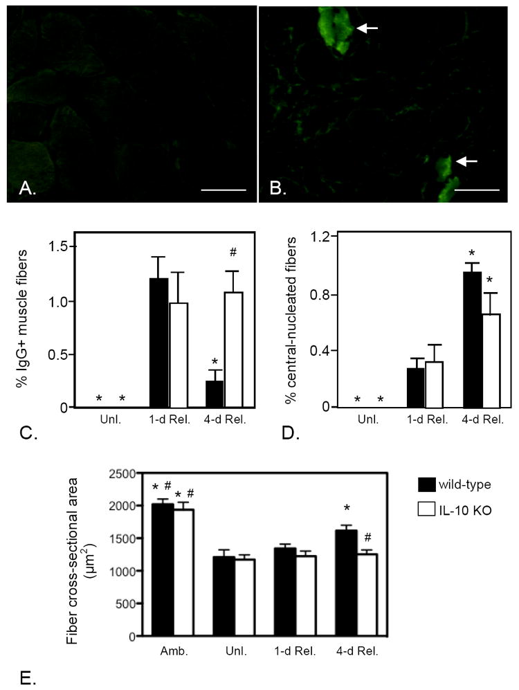
Figure 6.

IL-10 mutants experience reductions in muscle fiber repair, regeneration and growth during reloading. A, B: Cross-section of soleus muscles from wild-type (A) or IL-10 null mutant (B) mice from 4-days reloaded mice labeled with FITC-conjugated, anti-mouse IgG. The presence of IgG in the muscle fiber cytosol (arrows) indicates the presence of membrane lesions large enough to allow influx of IgG from the extracellular space. Bars = 100 μm. C: Quantification of the number of IgG+ muscle fibers in unloaded (Unl.), 1-day and 4-day reloaded muscles indicates that membrane lesions caused by muscle reloading are repaired between days 1 and 4 of reloading in wild-type, but not IL-10 mutant, muscles. D. Quantification of the proportion of central-nucleated muscle fibers in reloaded muscles shows that the increase in regenerative muscle fibers that occurs in wild-type muscles between in 1-day and 4-days reloading is diminished in IL-10 null mutants. E. Muscle fiber growth during reloading was assayed by measuring changes in cross-sectional area of the muscle fibers in soleus muscle sections. Note that fiber size in ambulatory controls (Amb.) did not differ between wild-type and IL-10 mutant muscles and that mutation of IL-10 did not affect the atrophy of fibers that occurred during unloading (Unl.). However, fiber growth that occurred during 4-days of reloading did not occur in IL-10 mutants. Each bar represents the mean and sem for the muscles collected from 6 mice in each data set. * indicates significantly different from 1-day reloaded muscle of the same genotype at p < 0.05. # indicates significantly different from 4-days reloaded wild-type muscle at p < 0.05.
