
Figure 7.
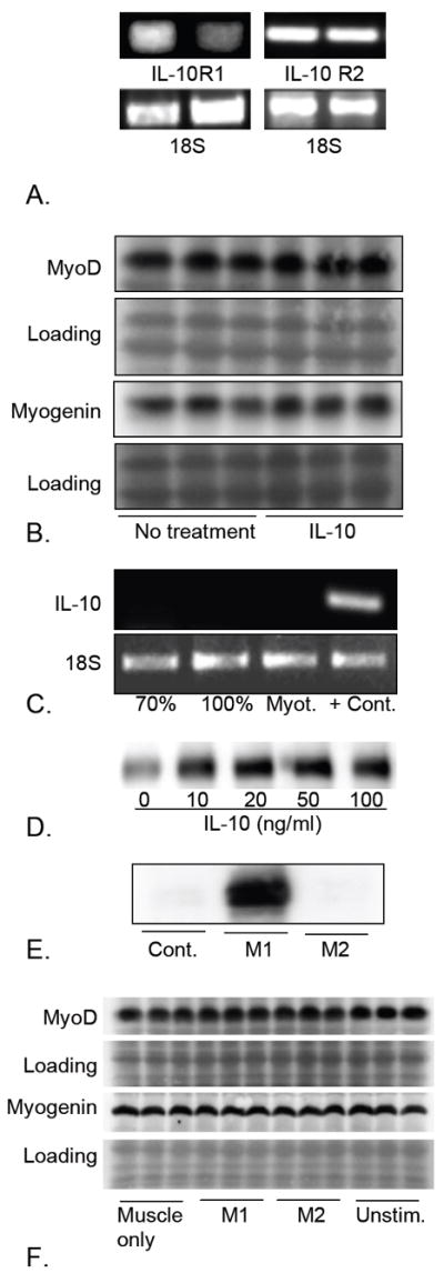
MyoD and myogenin expression are unaffected by direct stimulation with IL-10 or co-culture with IL-10 stimulated M2 macrophages. A. RT-PCR was used to confirm that C2C12 myoblasts can express both IL-10R1 and IL-10R2. The 18S ribosomal subunit was used as a loading control. B. Western blots of muscle cell extracts following treatment with 10 ng/mg IL-10 shows that direct application of IL-10 for 24 hours did not affect expression of MyoD or myogenin. Ponceau red staining of membranes that were subsequently used for antibody incubations was used to confirm uniform loading of the gels and transfer of proteins to the membrane (loading). The western blots that are shown are representative of three independent experiments. C: RT-PCR showed that muscle cells in vitro do not express IL-10, showing that lack of treatment effect with exogenous IL-10 was not attributable to saturation by endogenous IL-10. Lane 1 sample was obtained from 70% confluent cultures of proliferative muscle cells. Lane 2 was from 100% confluent cultures. Lane 3 was obtained from differentiated myotube (Myot.) cultures, 2-days after transfer to differentiation medium. Lane 4 used RNA isolated from inflamed, dystrophic muscle from mice in the mdx line, as a positive control. The 18S ribosomal subunit is used as a loading control. The results are representative of those obtained from 3 independent experiments. D. Western blot of extracts of macrophages stimulated with IL-10 for 24 hours. IL-10 induction of CD163 expression indicates increased activation to the M2 phenotype. The blot is representative of 3 independent experiments. E. Western blot of extracts of macrophages stimulated with TNF-α and IFNγ for 24 hours. Induction of iNOS indicates activation to the M1 phenotype. F. Western blot of extracts of myoblasts cultured in the absence of macrophages (Muscle only), or in the presence of M1 macrophages activated by TNF-α and IFNγ (M1), or in the presence of M2 macrophages stimulated with IL-10 (M2) or macrophages that were not treated with cytokines (Unstim.) No detectable changes in the levels of expression of MyoD or myogenin were observed in any co-culture conditions, compared to myoblasts alone. The results are representative of those obtained from 3 independent experiments. Ponceau red staining of the western blot membranes (Loading) was used to confirm uniform loading and transfer of samples.