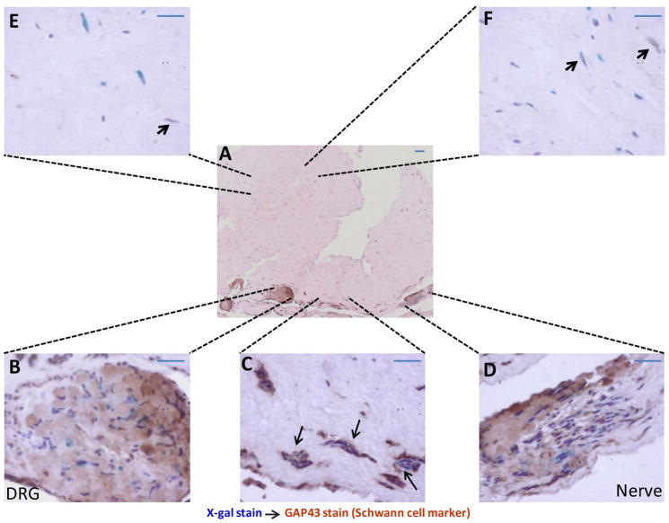
Figure 2. Nerve microenvironment promotes the differentiation of SKPs into Schwann cells.
In vitro engineered skin rafts containing lacZ-postive SKPs, fibroblasts and DRGs/nerves were harvested after 4–6 weeks in culture, using X-gal staining to trace the location of the SKPs. Tissues were then processed for histological and immunohistochemical analysis. The majority of blue cells (marker for SKPs) are also positive for GAP 43 (a Schwann cell marker) within and in the vicinity of the DRGs/nerves (A–D). There are tubal structures or cellular corridors of SKP-derived GAP 43 positive Schwann cells in these areas (C, arrows). In contrast, there are only a few LacZ-GAP 43 double positive cells in areas distant from the DRGs and nerves (E, F and arrows). Scale bar = 20 μm.

