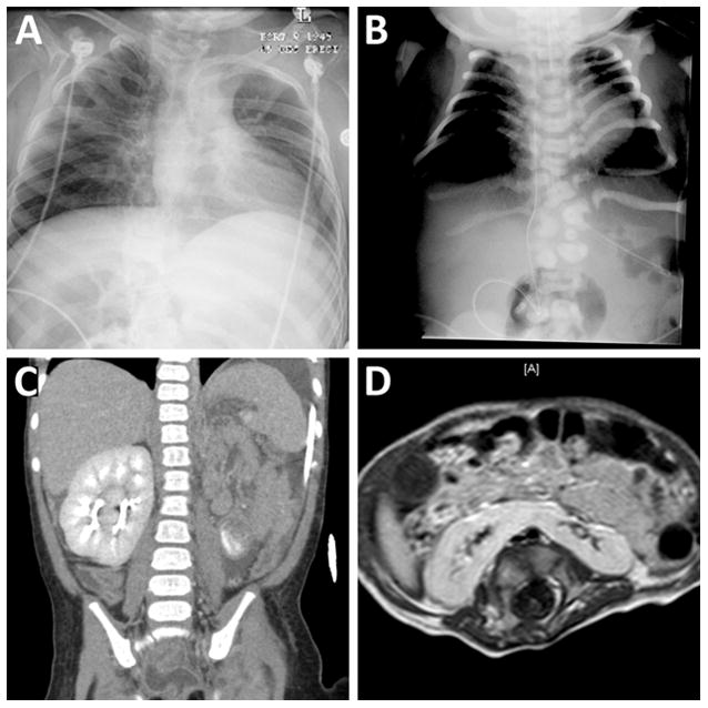
Figure 4. VACTERL Features.

A: Patient 33. Radiograph demonstrating multiple fused ribs associated with vertebral segmentation defects. B: Patient 35. Radiograph demonstrating multiple thoracic and lumbar vertebral segmentation defects and deformed ribs. C: Patient 21. Coronal CT showing absent kidney on the left with partially duplicated kidney on the right (cross-fused renal ectopia). D: Patient 36. Axial T1-weighted image of the abdomen showing horseshoe kidney.
