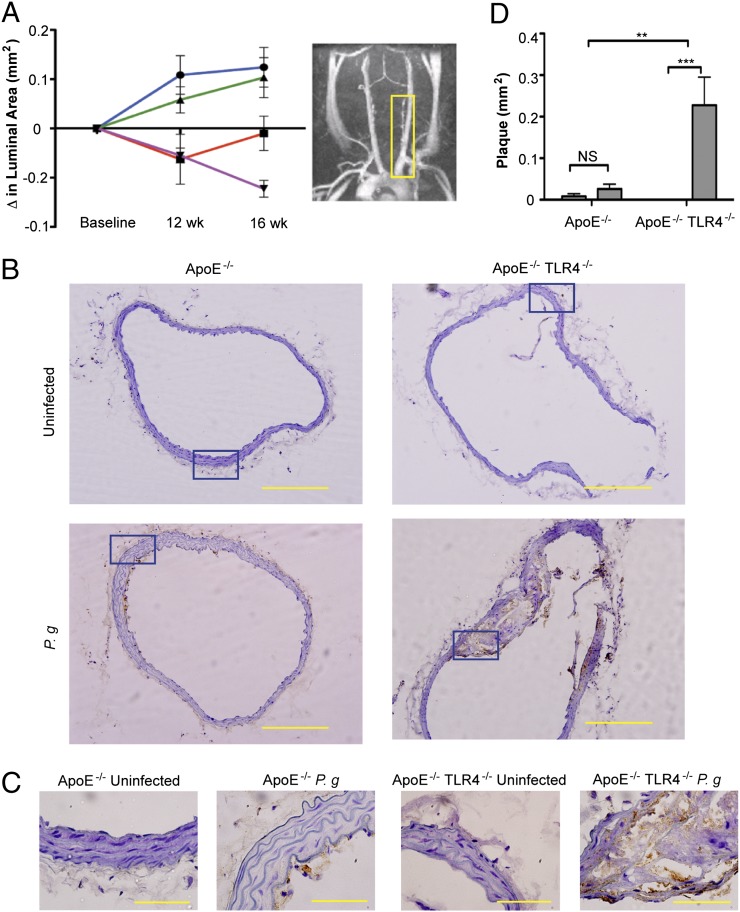
FIGURE 1.
TLR4 deficiency confers enhanced susceptibility to atherosclerosis in the innominate artery after infection with P. gingivalis. Innominate arteries were imaged by MRA at baseline (week 0) and at 12 and 16 wk after first oral infection. (A) The temporal change in luminal area (mm2) was calculated for individual mice (n = 5/group). Inset, Representative MRA image indicating the innominate artery (yellow box), where measurements were taken. Uninfected ApoE−/− (blue); P. gingivalis-infected ApoE−/− (red); uninfected ApoE−/−TLR4−/− (green); P. gingivalis-infected ApoE−/−TLR4−/− (purple). (B) Representative hematoxylin staining from each group in innominate artery with F4/80 staining (macrophages stain brown). Scale bar, 20 μm. (C) Visualization of intima, media, and adventitia of representative images. Areas indicated in (B) (blue box). Scale bar, 5 μm. (D) Plaque area within the innominate artery measured from histological images using IPLab software (Becton Dickinson) (n = 5/group). Black bar, Uninfected; gray bar, P. gingivalis infected. **p < 0.01, ***p < 0.001.

