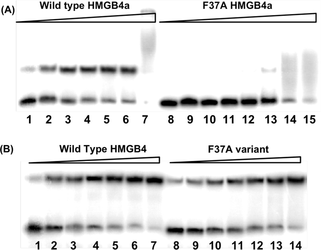
Figure 5.
EMSA analysis of HMGB4 protein binding to platinated DNA. (A) Wild type HMGB4a and its F37A variant bound to the cisplatin-modified DNA probe (1 nM). Protein concentrations were 2 nM (lanes 1, 8), 6 nM (lanes 2, 9), 20 nM (lanes 3, 10), 60 nM (lanes 4, 11), 200 nM (lanes 5, 12), 600 nM (lanes 6, 13), 2 µM (lanes 7, 14), and 6 µM (lane 15, F37A variant only). (B) Wild type and F37A variant of the full-length HMGB4 bound to the cisplatin-modified DNA probe (1 nM). Protein concentrations were 1 nM (lanes 1, 8), 2 nM (lanes 2, 9), 6 nM (lanes 3, 10), 10 nM (lanes 4, 11), 20 nM (lanes 5, 12), 60 nM (lanes 6, 13), and 100 nM (lanes 7, 14).

