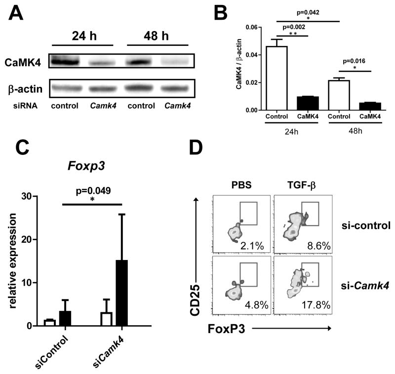
Figure 6. CaMK4 regulates FoxP3 in T cells from SLE patients.
A, T cells from 4 patients with SLE were transfected with either control siRNA or CAMK4-specific siRNA. A representative image of CaMK4 expression (western blot) at 24 and 48 hr after transfection is shown in the left panel. The right panel shows cumulative data from 4 experiments. B, 24 hr after transfection cells were either left unstimulated (white bars) or stimulated with anti-CD3 and anti-CD28 plus TGF-β (5 ng/mL; black bars). After 96 hr, FoxP3 expression was measured by real time PCR (left panel) or flow cytometry (right panel).

