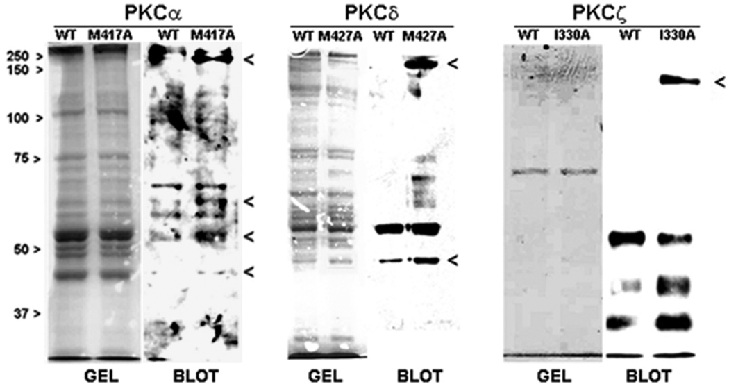
Figure 4.
Phospho-protein profiles of WT and traceable PKC isoforms. MCF-10A transfectants expressing FLAG-tagged WT or traceable mutant of each PKC isoform were lysed in detergent-free medium and each lysate was immunoprecipitated with anti-FLAG, as described in the ‘Methods’. This step facilitated the isolation of high affinity substrates of each PKC isoform. Following addition of N6-phenyl-ATP to each immunoprecipitated enzyme under activating conditions (phosphatidylserine for PKCα and δ, ceramide for PKCζ), phosphorylated products were resolved on SDS-PAGE and stained with Gelcode Blue. Western blot analysis of an aliquot of each sample (representing 25% of the total sample) was carried out with a duplicate gel and the resulting blot was probed with PKC substrates antibody (1:1000). Each WT/traceable mutant pair was aligned with the corresponding Gelcode blue-stained gel that serves as the loading control. Bands excised for MS/MS analysis are indicated with a caret (>). Each phospho-protein profile is representative of three independent experiments.

