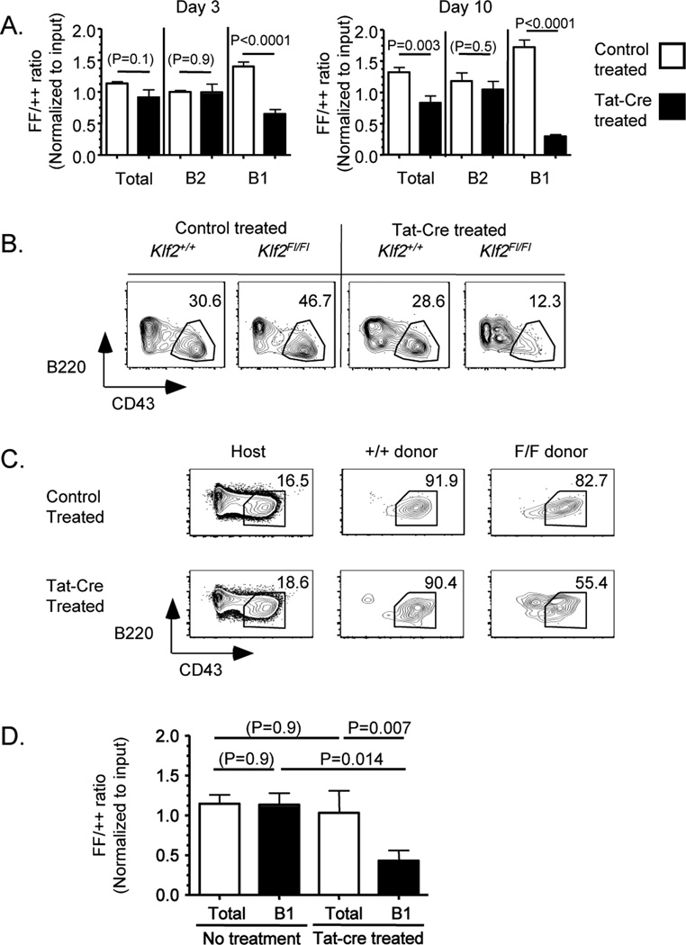
Figure 2. Induced KLF2 deletion in mature peritoneal B1 B cells leads to loss of B1 B cell phenotype.
In (A,B), peritoneal cells were isolated from congenic mice of either Klf2+/+ (++) or Klf2Fl/Fl (FF) genotype, and treated with Tat-cre prior to adoptive transfer. The data in (A) represent the ratio of Klf2Fl/Fl to Klf2+/+ donor cells, for the B cell subsets indicated, among the peritoneal lavage population at the time point indicated. These data are compiled from 3 experiments. (B) Representative B220/CD43 staining of the indicated donor populations from day 10 post-transfer. In (C,D) peritoneal B1 B cells were sorted from Klf2+/+ (++) or Klf2Fl/Fl (FF) mice and treated with Tat-cre or control prior to adoptive transfer. At day 10, cells were isolated from the peritoneum of the host. (C) Representative data for B220/CD43 expression on the Klf2+/+ and Klf2Fl/Fl donor populations (as well as host cells, for reference). In (D) the ratio of Klf2Fl/Fl to Klf2+/+ donor cells among the total B cell and B1 phenotype population is shown, for control and Tat-cre treated populations. Data were compiled from 3 experiments. (The number of animals represented are 9 mice per group in A; 3 untreated and 8 Tat-cre treated in D).

