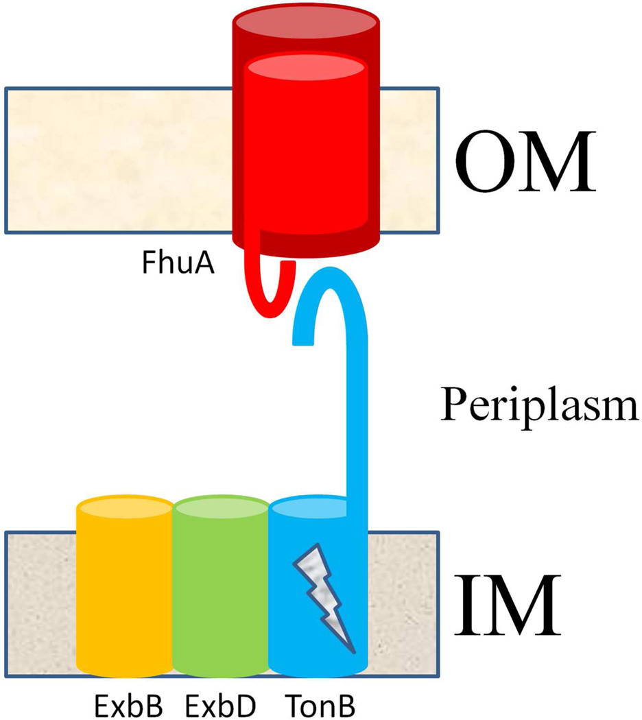
Figure 1.
Cartoon of spatial arrangement of the siderophore outer membrane permeation machinery. Siderophore receptors (FhuA is depicted in the figure) are TBDTs that sit in the outer membrane (OM) of gram negative bacteria and possess a conserved beta-barrel (dark red) architecture. Upon siderophore binding to the receptor, the plug domain of the receptor (light red) extends its TonB box (red hook), located near the amino terminus of the polypeptide, into the periplasm to interact with the carboxy terminus (blue hook) of the TonB protein of the inner membrane (IM) TonB complex, consisting of ExbB, ExbD and TonB. In an energy-dependent manner (indicated by a lightning bolt on TonB), TonB’s interaction with the receptor’s TonBbox somehow allows a pathway to form within the lumen of the receptor’s barrel, through which the siderophore diffuses.

