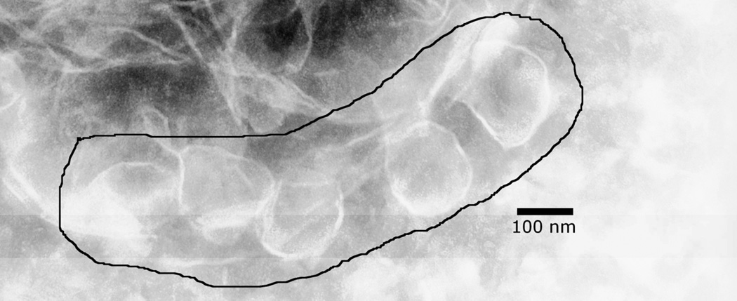
Figure 6.
FhuA forms vesicles in detergent solution. Shown here is a negative stained electron microscopy image at 50,000 magnification of whole FhuA after it had been diluted 10-fold into the buffer used in the bilayer experiments. (This resulted in a 0.03mg/mL FhuA protein concentration and a 0.01% LDAO detergent concentration.) The black line encloses several ~100 nm vesicles. No vesicles were observed in solutions with detergent alone (not shown).

