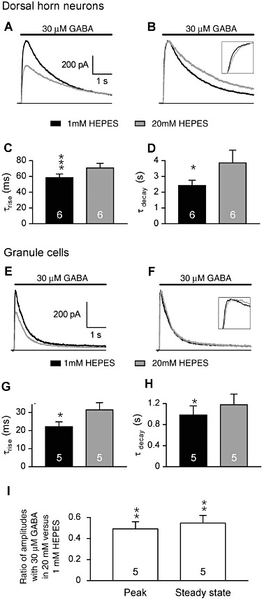
Figure 2.

HEPES slowed the kinetics of the currents induced by rapid applications of 30 µM GABA on spinal cord DH neurones and GCs. Patched neurones were detached from the dish and maintained close (∼100 µm) to the perfusion system. (A and B) The currents induced by 5 s applications of GABA (30 µM) in a DH neurone displayed slower rise and decay kinetics. Traces in A are normalized in B to compare kinetics. (E–F) The currents induced by 5 s applications of GABA (30 µM) in a GC displayed slower rise and decay kinetics. Traces in E are normalized in F to compare kinetics. (C and G) The average time constant values of the exponential functions fitting the current rise in DH (C) and GC (G) neurones significantly increased in 1 and 20 mM HEPES. (D and H) The average time constants of the exponential functions fitting the current decay in DH (D) and GC (H) neurones were significantly increased in 20 mM HEPES as compared with 1 mM HEPES. (I) Increasing HEPES from 1 to 20 mM similarly reduced the amplitude of the peak and of the steady state currents induced by 30 µM GABA in GC. All experiments were performed at pHe 7.3. Data in bar graphs are means ± SEM. *P < 0.05; **P < 0.01; ***P < 0.001; ratio values significantly different from unity; n.s., not significant; paired Student's t-test.
