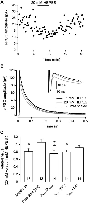
Figure 8.

The amplitude of electrically evoked GABAergic IPSCs (eIPSCs) is reduced by HEPES in DH neurones. (A) In a neurone recorded with 1 mM HEPES, the superfusion with an extracellular solution containing 20 mM HEPES induced a reversible decrease in the amplitude of GABAergic eIPSCs. (B) Superimposed eIPSCs recorded in 1 mM and 20 mM HEPES. The dark grey trace corresponds to the currents recorded in 20 mM HEPES that was normalized to the peak of the currents recorded in 1 mM HEPES. The inset shows details of the initial part of the traces. The stimulus artefact was blanked for 20 mM HEPES traces. (C) Average values of GABAergic eIPSC amplitude and kinetic properties recorded with 20 mM HEPES, normalized to the values recorded in presence of 1 mM HEPES. All experiments were performed at constant pHe 7.3. Data represent the mean of normalized values ± SEM. *P < 0.05; **P < 0.01; ratio values significantly different from unity; n.s., not significant; paired Student's t-test.
