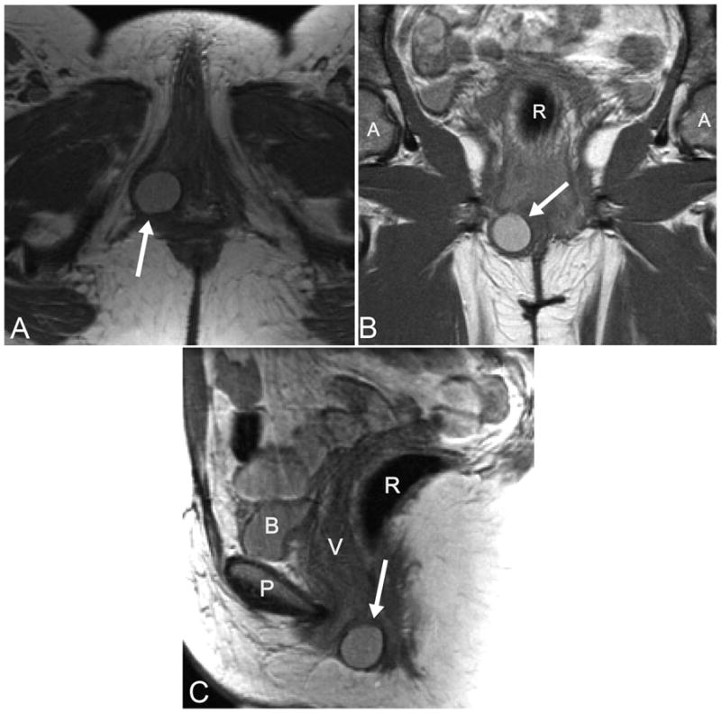
Figure 1.

A: Axial slice of a proton-density magnetic resonance imaging (MRI) scan demonstrating a Bartholin gland cyst (arrow).B: Coronal slice of an MRI scan with a visible Bartholin gland cyst (arrow). C: Sagittal slice of an MRI scan with a Bartholin gland cyst identified (arrow). R, rectum; A, acetabulum; P, pubic symphysis; B, bladder; V, vagina.
