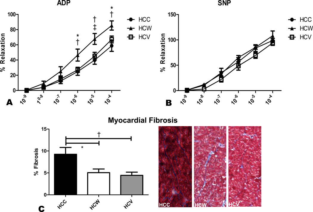
Figure 4. Microvessel function and myocardial fibrosis.
Microvessel relaxation responses to endothelium-dependent ADP and endothelium-independent SNP were measured in coronary arterioles from the AAR. A significant improvement in endothelium-dependent vasodilation was seen in the HCW group compared to both HCC and HCV (A). There was no difference between groups in endothelium-independent vasodilation (B). Myocardial fibrosis as measured by trichrome staining was significantly higher in the HCC group than in both other groups. Shown are representative trichrome stained myocardial sections from the three groups, with myocardium staining red and intervening connective tissue staining blue (C). *p<0.05, †p<0.01, ‡p<0.001.

