Figure 5.
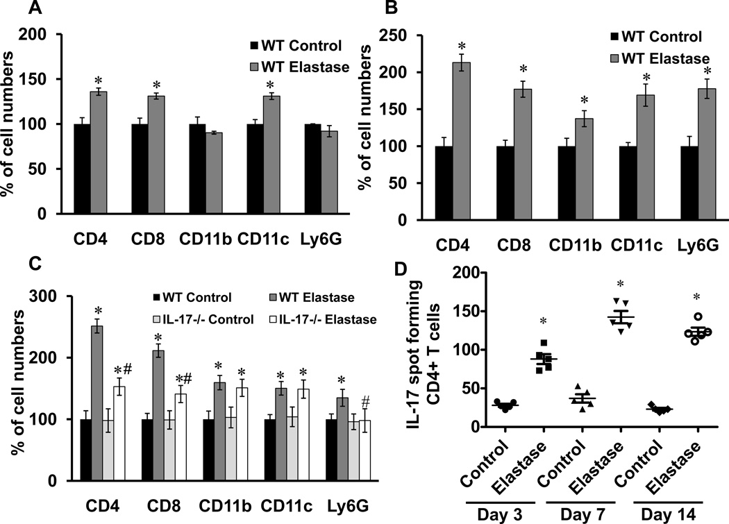
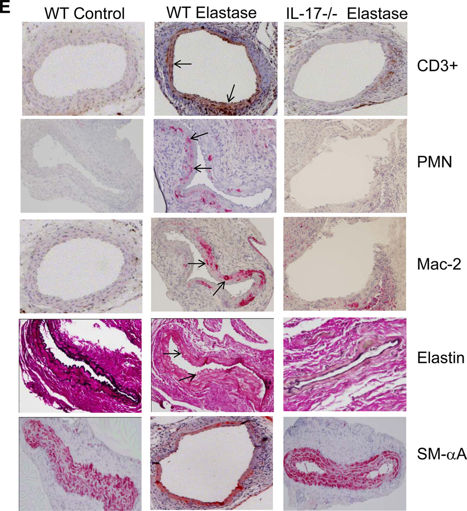
CD4+ T cell infiltration and IL-17 production is increased in aortas from elastase-perfused WT mice. Flow cytometry analysis of cell counts was performed on aortic tissue from control or elastase-perfused WT mice on days 3, 7 and 14. Results are shown as percentage of cell numbers compared to WT controls; n=4–5 mice/group; *p<0.05 vs. WT control, #p<0.05 vs. WT elastase. A. On day 3, a significant increase in CD4+ and CD8+ T cells as well as CD11c+ dendritic cells was observed in elastase-perfused WT mice compared to controls. B. On day 7, a significant increase in CD4+ and CD8+ T cells, CD11b+ (macrophages), CD11c+ (dendritic) and Ly6G+ (neutrophils) was observed in elastase-perfused WT mice compared to controls. C. On day 14, a significant increase in all cell populations remained significantly increased in elastase-perfused WT mice compared to controls. Elastase-perfused IL-17−/− mice had a significant attenuation in CD4+ and CD8+ T cells as well as Ly6G+ neutrophils compared to elastase-treated WT mice. D. ELISPOT analysis of IL-17-producing spot forming CD4+ T cells demonstrated a significant increase in CD4+ T cell-producing IL-17 cells in elastase-perfused aortic tissue from WT mice compared to controls on days 3, 7 and 14. E. Comparative histology and immunohistochemistry performed on day 14 indicates a significant decrease in CD3+ T cell, neutrophil (PMN) and macrophage (Mac-2) immunostaining, increase in smooth muscle cell α-actin (SM-αA) expression (arrows indicate areas of immunostaining), and decrease in elastic fiber disruption in aortic tissue (Verhoeff-Van Gieson staining for elastin) of elastase-perfused IL-17−/− mice compared to elastase-perfused WT mice. n=3–5/group; *p<0.05 vs. WT control.


