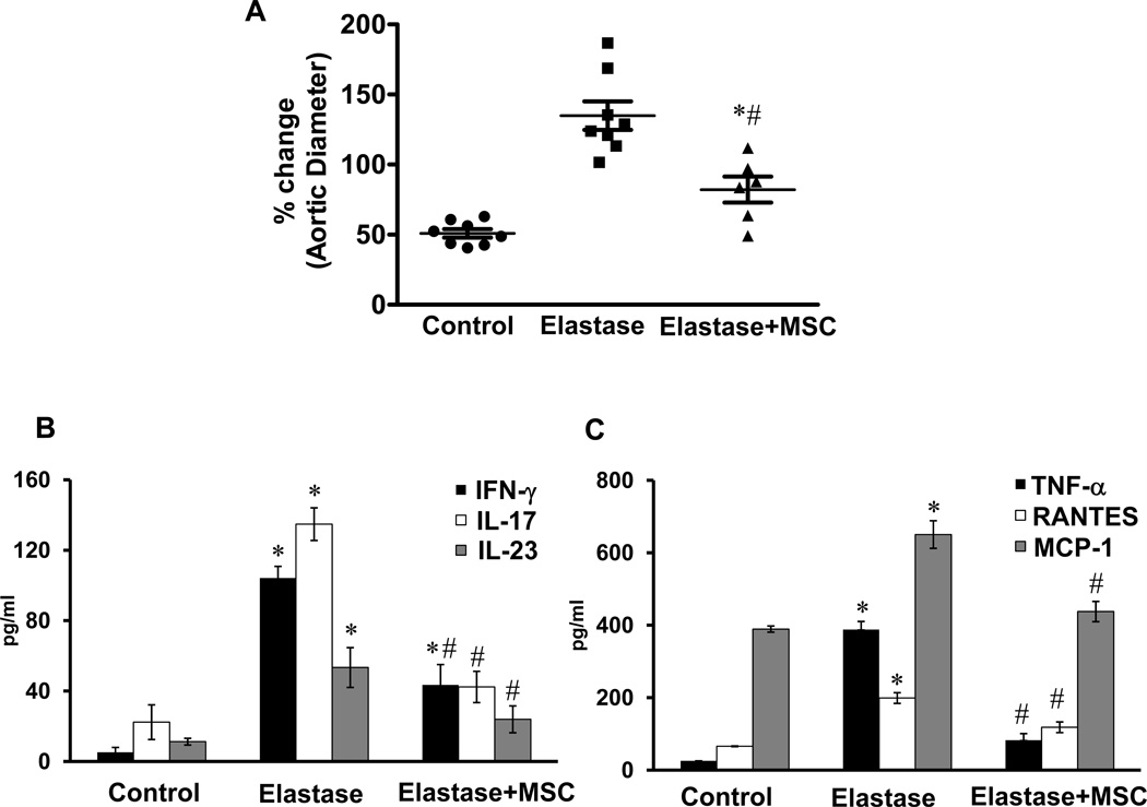
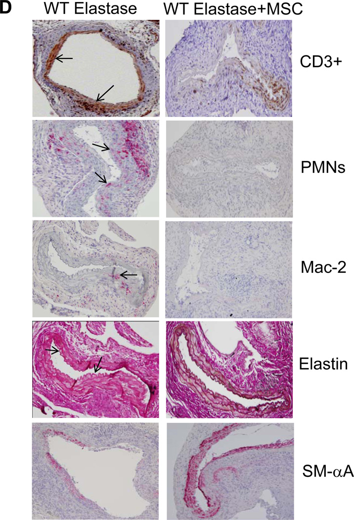
Figure 8.
MSCs attenuate AAA formation. A. Aortic diameter measurement of elastase-perfused WT mice treated with MSCs showed a significant attenuation in AAA formation compared to elastase-perfused WT mice alone on day 14. B. Aortic tissue from elastase-perfused WT mice treated with MSCs showed a significant attenuation in IL-17 and IL-23 production compared to elastase-perfused WT mice alone. C. MSC treatment significantly attenuated pro-inflammatory cytokine/chemokine production in elastase-perfused WT mice. D. Comparative histology and immunohistochemistry performed on day 14 indicates that WT mice treated with MSCs have a significant decrease in CD3+ T cell, neutrophil (PMN) and macrophage (Mac-2) infiltration (arrows indicate areas of immunostaining), increase in smooth muscle cell α-actin (SM-αA) expression as well as decrease in elastic fiber disruption (Verhoeff-Van Gieson staining for elastin), compared to elastase-perfused WT mice. n=5–9 mice/group; *p<0.05 vs. control; #p<0.05 vs. elastase.


