Figure 3.
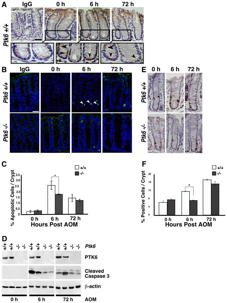
Induction of PTK6 in colonic crypts following AOM treatment correlates with increased apoptosis and proliferation. (A) PTK6 immunohistochemistry was performed on Ptk6 +/+ colons at 6 or 72 hours after a single injection of AOM. Colon sections were counterstained with hematoxylin. The size bar represents 50 μm. Magnification of boxed areas (top panel) are shown below. Arrows denote PTK6 positive cells. (B) Cleaved Caspase-3 immunohistochemistry was performed on colon sections from Ptk6 +/+ and Ptk6 −/− mice sacrificed at 6 or 72 hrs post AOM injection. Size bar represents 50 μm. (C) Cleaved Caspase-3 positive cells were counted in ten representative colonic crypts. Increased numbers of cleaved Caspase-3 positive cells were detected at 6 hours post AOM injection in Ptk6 +/+ mice (* P < 0.05, Bars +/− SD). (D) Immunoblotting was performed with lysates harvested at 6 or 72 hours post AOM injection, using antibodies directed against PTK6, cleaved Caspase-3, and β-actin. (E) BrdU immunohistochemistry was performed on Ptk6 +/+ and Ptk6 −/− mice sacrificed at 6 or 72 hours post AOM injection. BrdU was injected 2 hours before animals were killed. (F) BrdU positive cells were counted in ten representative colonic crypts in Ptk6 +/+ and Ptk6 −/− mice. The number of BrdU positive cells is higher in Ptk6 +/+ mice (* P < 0.05, Bars +/− SD).

