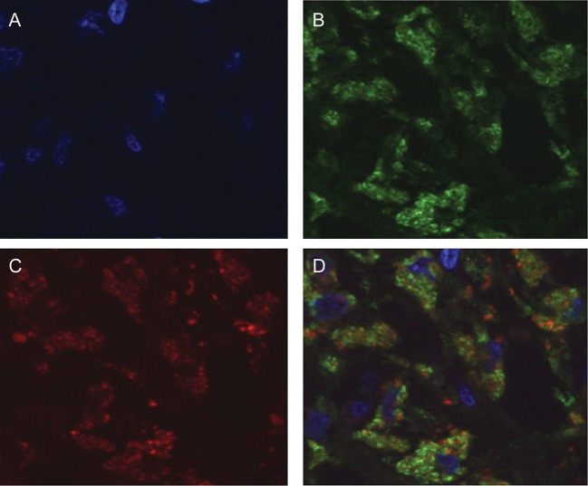
Figure 3.
CD68 and interleukin 17 (IL-17) colocalization in macrophages in lymph node tissues from patients coinfected with human immunodeficiency virus-1 (HIV-1) and Mycobacterium avium complex (MAC). Lymph node tissue sections were stained by indirect immunofluorescence, using both mouse anti-CD68 and rabbit anti–IL-17 antibodies, followed by corresponding secondary antibodies conjugated to either Alexa-488 or Alexa-546. Sections were also stained with Hoechst. A, Nuclear detection as determined by Hoechst staining (blue). B, Macrophages in coinfected lymph nodes show positive immunofluorescence staining for CD68 (green). C, The same section shown in A and B is also positive for IL-17 (red). D, Overlay images (A, B, and C) demonstrate colocalization of CD68 and IL-17 in macrophages of coinfected lymph nodes. Original magnification is 100×.

