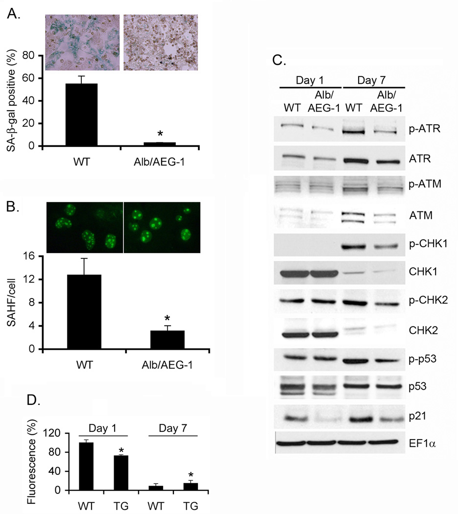
Fig. 4. Alb/AEG-1 hepatocytes are resistant to induction of senescence.
A. Photomicrograph of WT and Alb/AEG-1 hepatocytes stained for senescence-associated β-galactosidase (SA-β-gal) after 1 week of culture. Graphical representation of quantification of SA-β-gal positive cells. B. Photomicrograph of WT and Alb/AEG-1 hepatocytes stained for γ-H2AX. Graphical representation of quantification of γ-H2AX foci/cell. For A and Bthe data represent mean ± SEM of three independent experiments. *: p<0.01. C. Analysis of expression level of the members of DNA damage response pathway in WT and Alb/AEG-1 hepatocytes by Western blotting. D. Total ROS level in WT and Alb/AEG-1 (TG) hepatocytes at day 1 and day 7 of culture. The data represent mean ± SEM of four independent experiments. *: p<0.01.

