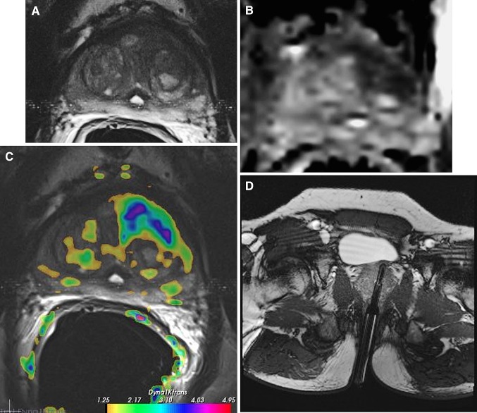
Fig. 1.
Multi-parametric MR images, comprising T2-weighted (A), diffusion weighted-derived apparent diffusion coefficient map image (B), and dynamic contrast-enhanced image (Ktrans) (C), in a 63 year old male, with a PSA of 12 ng/ml, and a history of two negative transrectal ultrasound guided prostate biopsy sessions. In the left transitional zone a cancer suspicious region is present (low-signal intensity lesion on T2, asymmetric Ktrans and restricted diffusion on the ADC map). D During a second session, a MR-guided biopsy was performed of the cancer suspicious area. The needle guider was pointed toward the cancer suspicious region in axial and subsequently biopsied. Verification image with the needle guider left in situ was obtained. Histopathology revealed a Gleason 4 + 3 prostate cancer.

