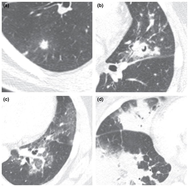
Figure 1.

Axial CT images demonstrating (a) nodule with a halo of surrounding GGO, (b) nodule with ‘air crescent’ sign of cavitation, (c) geographic areas of GGO and (d) multifocal parenchymal opacification ranging from ground-glass to consolidation. CT, chest computed tomography; GGO, ground glass opacity.
