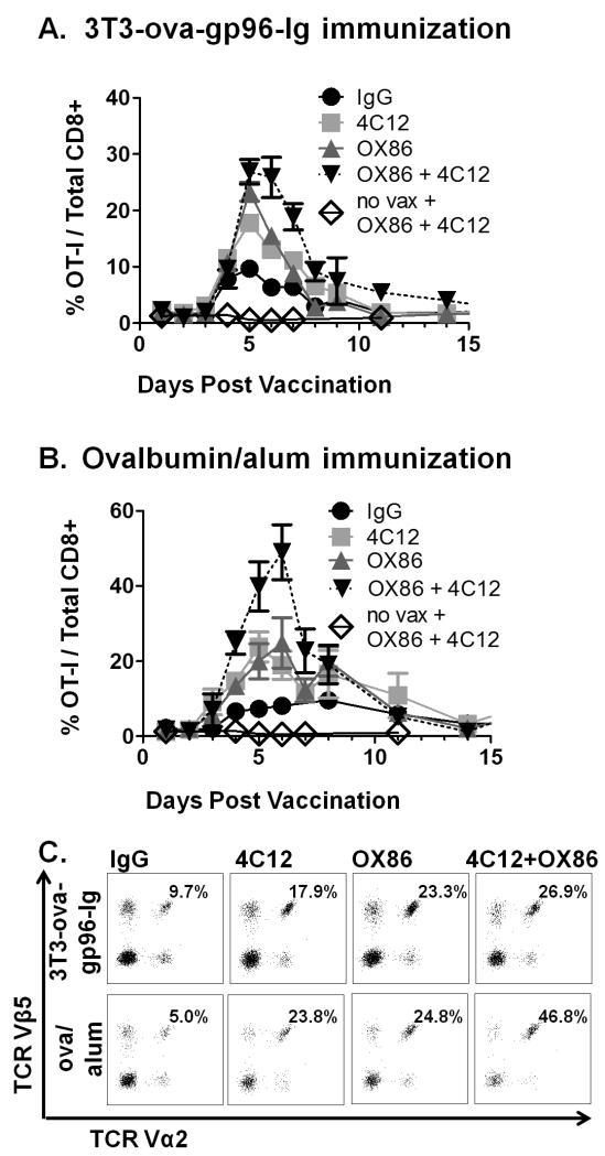
Figure 2. Comparative costimulation of OT-I proliferation by TNFRSF4 and TNFRSF25 agonistic antibodies.
Adoptive transfer of OT-I and OT-II cells was performed as described in figure 1, followed by immunization with either 3T3-ova-gp96-Ig (A) or ova/alum (B) together with either IgG control antibody (100 μg, black circles), TNFRSF25 agonistic antibodies (clone 4C12, 20 μg, red squares), TNFRSF4 agonistic antibodies (clone OX86, 100 μg, green triangles) or both 4C12 and OX86 antibodies combined (blue squares). OT-I proliferation in the peripheral blood was measured by flow cytometry on the indicated days, and representative plots of Vα2+Vβ5+ cells (pre-gated on CD3+CD8+ cells) are illustrated in (C) on day 5 for each condition. Data indicate mean ± SEM for ≥ 5 mice in each of ≥ 2 independent experiments.

